Women's Imaging
Sonohysterogram

What is a Sonohysterogram?
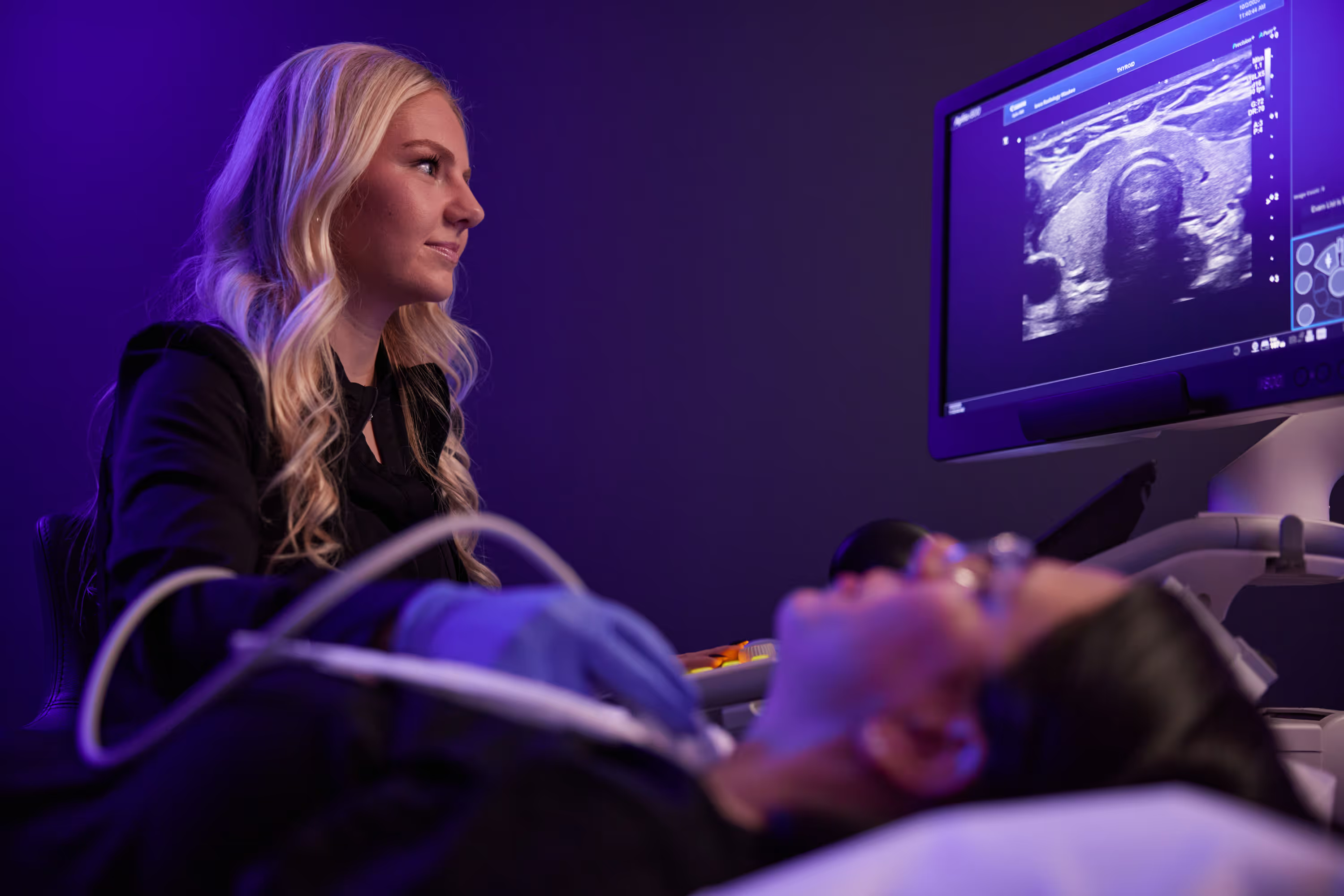
A sonohysterogram is an ultrasound procedure used to diagnose uterine and endometrial abnormalities. It can help to pinpoint areas of concern for abnormal uterine bleeding, fibroids, or polyps. Ultrasound does not use radiation. Ultrasound images are used to show the structure of the uterus and endometrium and its thickness.

What happens during the test?
Our technologist will take a brief medical history, and you will be asked to disrobe from the waist down and put on a gown. You will be asked to lie down face up on the examination table. The radiologist will insert a speculum into your vagina to access the cervix. The cervix will be sterilized, and a flexible catheter will be passed through the opening of the cervix. The speculum will then be removed, and a vaginal ultrasound camera (probe) will be inserted into the vagina.
The ultrasound probe will take pictures of the uterus, measuring endometrial thickness and shape. Sterile saline will be injected into the uterus, enlarging the cavity and outlining the lining for easy visualization. After several images are obtained, the probe will be removed. Cramping, similar to that experienced during menses, is common after the exam.
This exam typically takes 60 minutes to complete.
How do I prepare for the test?
Wear a loose-fitting two-piece outfit. Timing of your exam is essential and should be performed 4 to 8 days after the beginning of your period. Cramping is common, so we recommend taking up to 600 milligrams of ibuprofen 30 minutes before the exam.
When can I expect the results?
Our radiologist will discuss the results with you following your exam. In addition, he or she will analyze the images and send a signed report to your referring physician within 1 business day.
After the test
You may experience cramping and/or spotting and a watery discharge for one to two days following the procedure.
Downloadable Resources
FAQ's
Where we offer
Sonohysterogram

3625 N Ankeny Blvd. Suite H Ankeny, IA 50023
Mon – Fri: 8am-5pm
Sat: Hours available for MRI and Digital Mammography
Fax: (515) 963-7619

12368 Stratford Drive Suite 300 Clive, IA 50325
Mon – Fri: 6:30am–5pm
Sat: Hours available for MRI & Digital Mammography
Fax: (515) 226-8408
Sarah Agan, R.T. (R) (M)
Phone: 515-226-9810
Fax: 515-226-8408
Email: sagan@iowarad.com

1221 Pleasant Street Suite 350 Des Moines, IA 50309
Mon – Fri: 8am-5pm
Fax: (515) 226-7493

2515 Grand Prairie Parkway, Waukee, IA 50263
Mon – Fri: 8am-5pm
Fax: (515) 226-8408
Schedule Today
At Iowa Radiology, we strive for excellence in everything we do. You can expect easy access, convenient scheduling, and timely service.
Featured Articles
Schedule an Appointment
At Iowa Radiology, we strive for excellence in everything we do. You can expect easy access, convenient scheduling, and timely service.


